Acquisition and Data Requirements
Image Data |
Dynamic cardiac H215O PET study (decay corrected). |
TAC 1 |
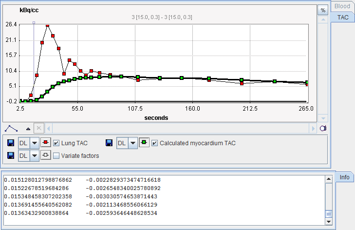
Time-activity curve representing blood activity in the lungs. |
Model Preprocessing
The lung TAC must be specified as a VOI or a file. It is then used together with the input parameters to calculate the expected TACs in the left ventricle (LV), the right ventricle (RV), and the myocardium.

Delay Lung-RV |
Left shift of the lung TAC to the time when the bolus arrived in the RV. |
Delay Lung-LV |
Right shift of the lung TAC to the time when the bolus arrived in the LV. |
Delay Lung-Myo |
Right shift of the lung TAC to the time when the bolus arrived in the myocardium. |
Mean Perfusion |
Expected mean perfusion of the myocardium. |
Partition Coefficient |
Partition coefficient of water in myocardium |
The results of preprocessing is shown in the Results panel.

Map Parameters

Myo |
The myocardium factor images which should represent an anatomical image of myocardium. |
BV |
Blood volume factor images which should show the blood volume. |