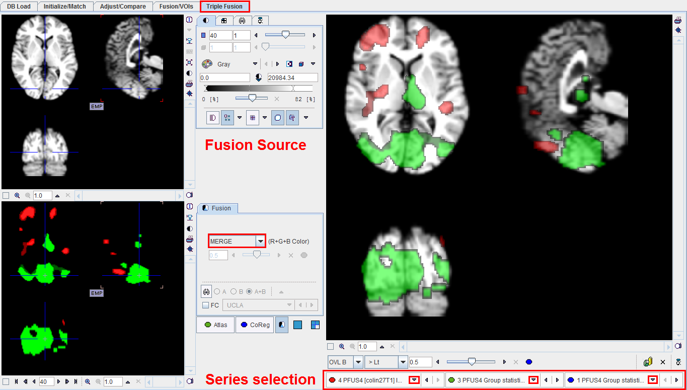
In some situations it may be helpful to compile the information of three images into one fused rendering. This can be done on the Triple Fusion page as illustrated below with a functional MRI study. Three studies were loaded, an anatomical data set and two contrasts. The series selection can be perform as indicated below.

The image selection  corresponds to the fusion source panel in the upper left. The next selections correspond to the Atlas and CoReg and are displayed in the CoReg panel in the lower left.
The anatomical MRI image was selected as fusion source. One contrast was selected as Atlas and the color table set to Red. The other contrast was selected as CoReg and the color table set to Green. The CoReg panel shows the fusion of the two image contrast, Atlas and CoReg using the MERGE mode.
The large image display to the right always shows the fusion of the image selected as fusion source with the image shown in the lower left part. So if the images are selected as in the example, the 3 source fusion shows the two contrasts on the anatomical image. In this configuration, the OVL B method was used to clearly see the anatomy outside the contrast area.
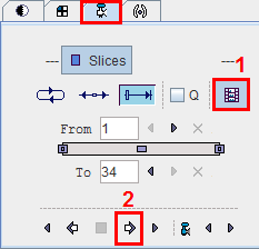
An additional helpful feature is the ability to capture the images in the right display into a sequence of JPEG files and to generate a movie. As soon as the Movie control button  in the lower right corner is activated all studies are switched into the Movie mode. Activate the Save video capture button 1 and starts with button 2 stepping the fusion images through all the slices of the currently selected plane. At the end the program prompts for a movie file name.

Select the plane around the vertical axis of which the projection should be rotated, and then start processing.
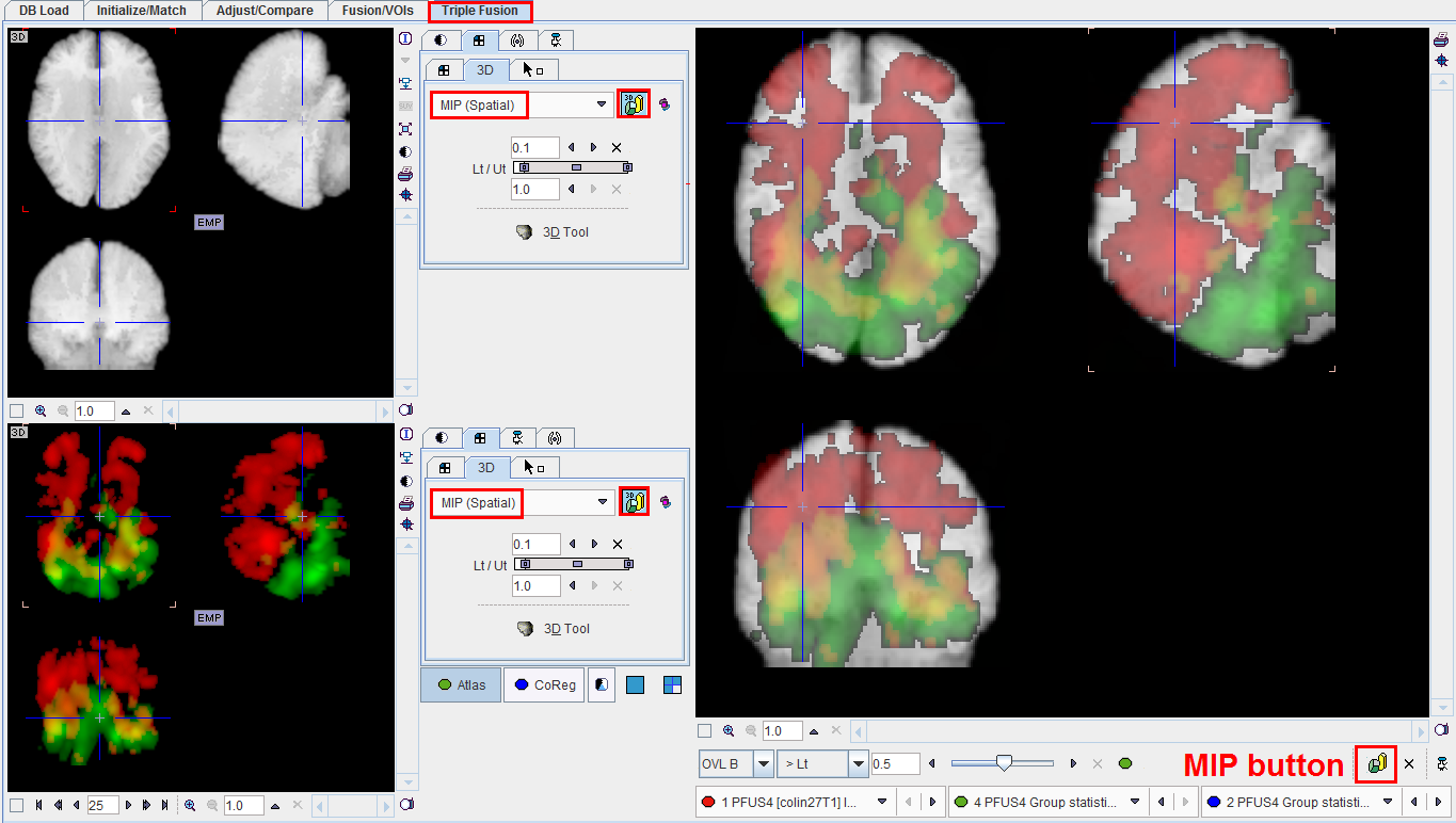
As soon as the MIP button is selected, all studies are switched into MIP mode:

Use the ![]() button to switch off the MIP mode.
button to switch off the MIP mode.