![]()
![]()
![]()
![]()
|
|
|
|
|
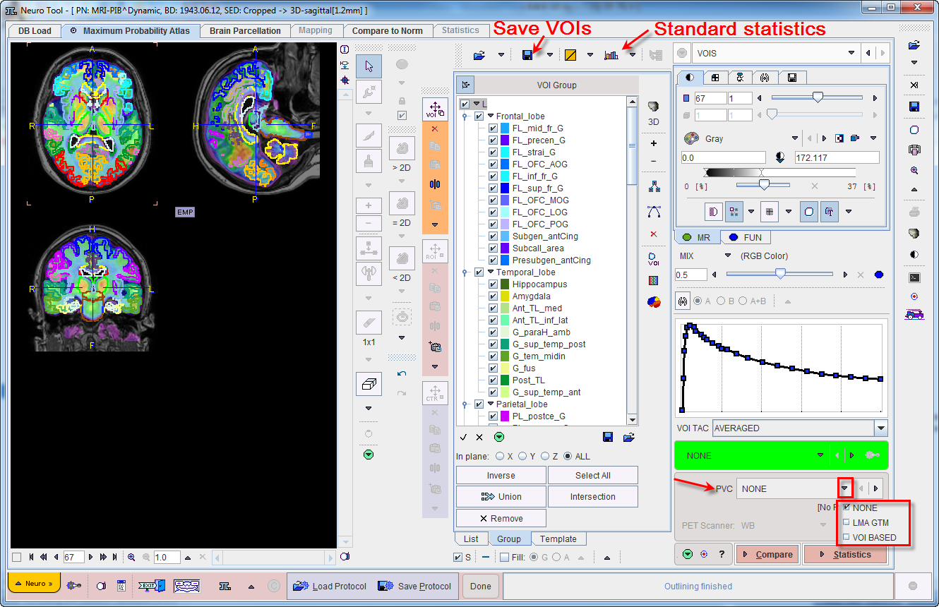
The result of structure outlining is shown on the VOIS page.
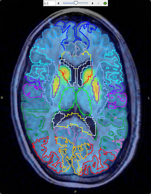
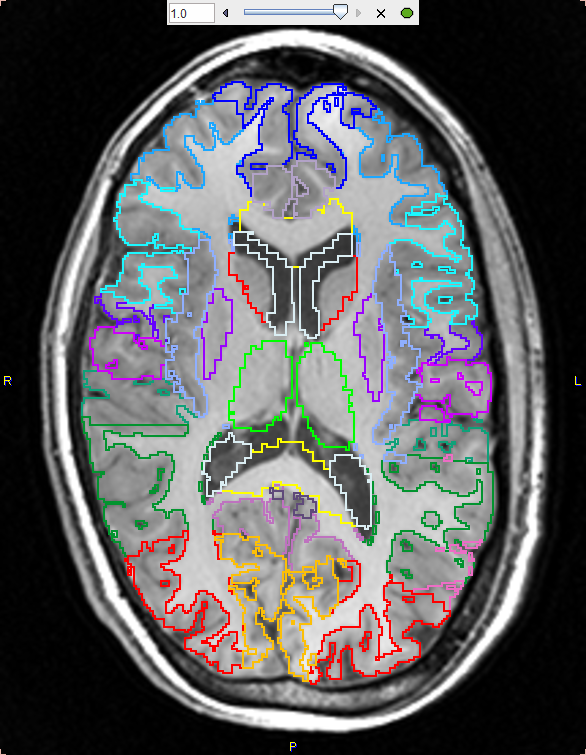
The image display shows a fusion of the MR image with the averaged PET image in the selected Result space. Please use the fusion slider to change the weight between the image contributions, and use the individual image control tabs for the changing the individual image displays. The example below shows the contour VOIs with 50% mixing and MR only.
VOI Editing and Selection
At this time the contour VOIs can be interactively adjusted using the VOI features of PMOD, which are described in the PMOD Base Functionality Guide. Note that the List tab should be selected for the adjustment, and that depending on the configuration only a reduced set of VOI tools may be available.
A subset of the VOIs can easily be selected on the Group tab as described above.
Partial-Volume Correction (PVC) Option
The PNEURO tool supports the GTM-based partial-volume correction (PVC) of the PET signal.
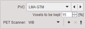
The PVC selection has three choices:
For the purpose of PVC, the VOIs complementary to the gray matter masked VOIs are taken into account. Also, all VOIs are considered, even if some were deleted by the user.
If a PVC method is used, both the original and the corrected statistics are calculated. Note that due to the high number of VOIs the PVC calculation may take several minutes and consumes a significant amount of RAM.
Statistics Calculation
Once the VOIs are acceptable, it is recommended to first save them and then proceed with statistics calculation by the Statistics action button. The result (of the selected VOIs only) is shown on the separate Statistics page of the PNEURO tool, from where it can be further evaluated.
Parametric Mapping
If the PET images are dynamic and the PXMOD option is included in the license, parametric mapping using pixel-wise models can be directly applied within PNEURO, as described in a separate section.
Normal Database Comparison
As PNEURO is able to provide the PET images normalized to the atlas space, there is a direct link with the Brain Norm Functionality. The Compare button copies the PET images as they appear on the VOIs page to the Compare To Norm page where they can be compared against a normal uptake pattern of a normal database. Conveniently, if the two spaces match, there is no need to perform a normalization.